Inner collateral ligament rupture

Anatomy

The bones of the knee joint include the femur (thigh bone), tibia (shin bone) and patella (kneecap). The knee joint is reinforced by a joint capsule that is reinforced with an external and internal collateral ligament (ligamentum collaterale laterale and mediale). Inside the knee are two ligaments, the anterior and posterior cruciate ligaments (ligamentum cruciatum anterius and ligamentum cruciatum posterius). The cruciate ligaments attach to the femur and tibia and hold these bones together, supporting and stabilising the knee. The anterior cruciate ligament prevents the lower leg from sliding forwards in relation to the femur.

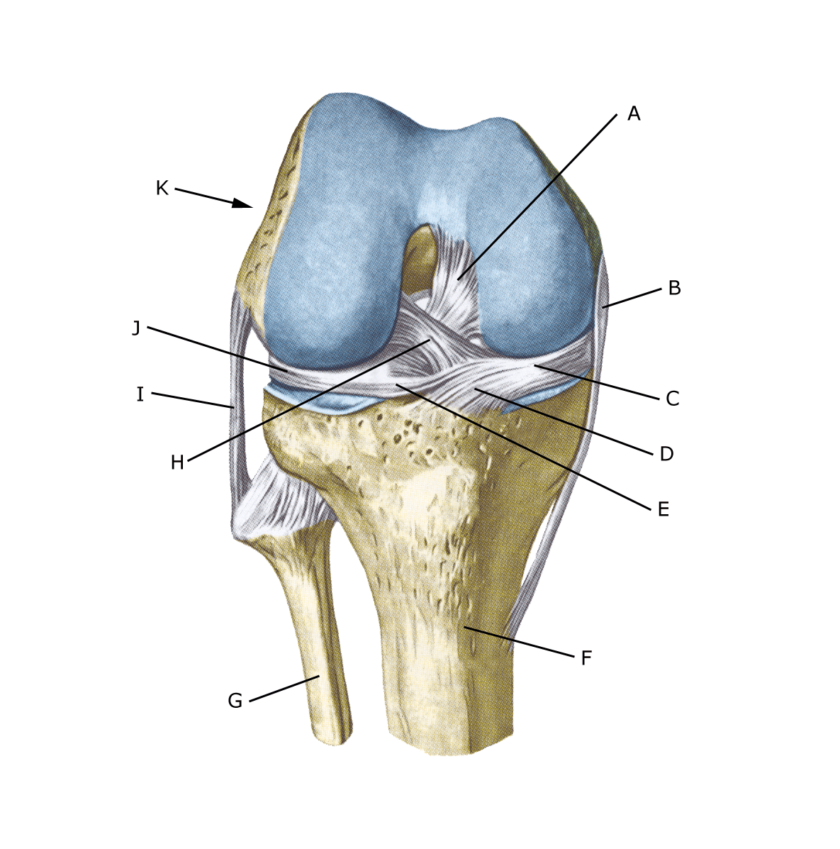
Knee joint from the front

A. Ligamentum cruciatum posterius
B. Ligamentum collaterale mediale/tibiale
C. Meniscus medialis
D. Insertio anterior menisci medialis
E. Ligamentum transversum genus
F. Tibiae
G. Fibulae
H. Ligamentum cruciatum anterius
I. Ligamentum collaterale laterale/fibulare
J. Meniscus lateralis
K. Femur

Cause

Anterior cruciate ligament rupture is a serious injury that usually occurs when the knee is twisted during football, handball, basketball, skiing or jumping gymnastics. The incidence is 6-8 times higher during matches compared to training, and girls are affected 50% more frequently than boys (Bram JT, et al. 2021).

Most injuries occur without contact with the opponent. ACL injuries in children are diagnosed with increasing frequency and only about half return to pre-injury levels (Sun B, et al. 2024).

Symptoms

Usually a pop is heard or felt and further sports activity must be cancelled. The knee swells within the first few hours, after which the knee cannot fully bend. There is often a sensation of the leg giving way (knee failure).

Examination

If you suspect a total or partial rupture of the anterior cruciate ligament, you should see a doctor (emergency room) immediately to make a diagnosis. The doctor can perform various knee tests (anterior drawer looseness, Lachmann, Pivot shift) to examine the stability of the knee. Please note that looseness in the knee can often only be detected after 2 weeks.

The diagnosis of a torn ACL usually requires an MRI scan. A 2-plane knee X-ray is also advised (within 2 weeks) if any of the following are present: the patient is under 12 years old, cannot walk 4 steps due to pain, pressure tenderness of the patella or fibular head (caput fibulae), the knee cannot bend more than 60°, or there is ligament damage (Pittsburgh knee rules).

Treatment

Pre-teenagers are usually reluctant to undergo surgery and therefore treatment is rehabilitation. If there has been knee failure or if the ACL injury is combined with other ligament or meniscus injuries, surgery with insertion of a ‘new’ ACL is recommended.

It takes about 9 months before children can return to sports after ACL surgery (Lorange JP, et al. 2024).

Rehabilitation

You should expect an intensive rehabilitation programme over a year. It is important to fully extend the knee at least twice a day. If there are problems with fully extending the knee, the operator should inspect the knee.

Bandage

The hinge bandage (Don-Joy) can be used for the first few weeks. Tape treatment for ACL ruptures in the knee has no certain effect.

Complications

If no progress is made, various complications must be considered.

In particular, the following should be considered:


In cases where the patellar tendon has been used as a new cruciate ligament, inflammation (‘inflammation’) is frequently seen in the patellar tendon where the patellar tendon has been removed. In children, there is a risk of affecting the growth of the operated leg as the growth zone must be pierced to insert the ACL.


As there is a risk of permanent damage, the injury should be reported to your insurance company.

Rehabilitation

Rehabilitation program