Rupture of the posterior cruciate ligament

Anatomy

The bones of the knee joint include the femur (thigh bone), tibia (shin bone) and patella (kneecap). The knee joint is reinforced by a joint capsule that is reinforced with an external and internal collateral ligament (ligamentum collaterale laterale and mediale). Inside the knee are two ligaments, the anterior and posterior cruciate ligaments (ligamentum cruciatum anterius and ligamentum cruciatum posterius). The cruciate ligaments attach to the femur and tibia and hold these bones together, supporting and stabilising the knee. The posterior cruciate ligament prevents the lower leg from sliding backwards in relation to the femur.

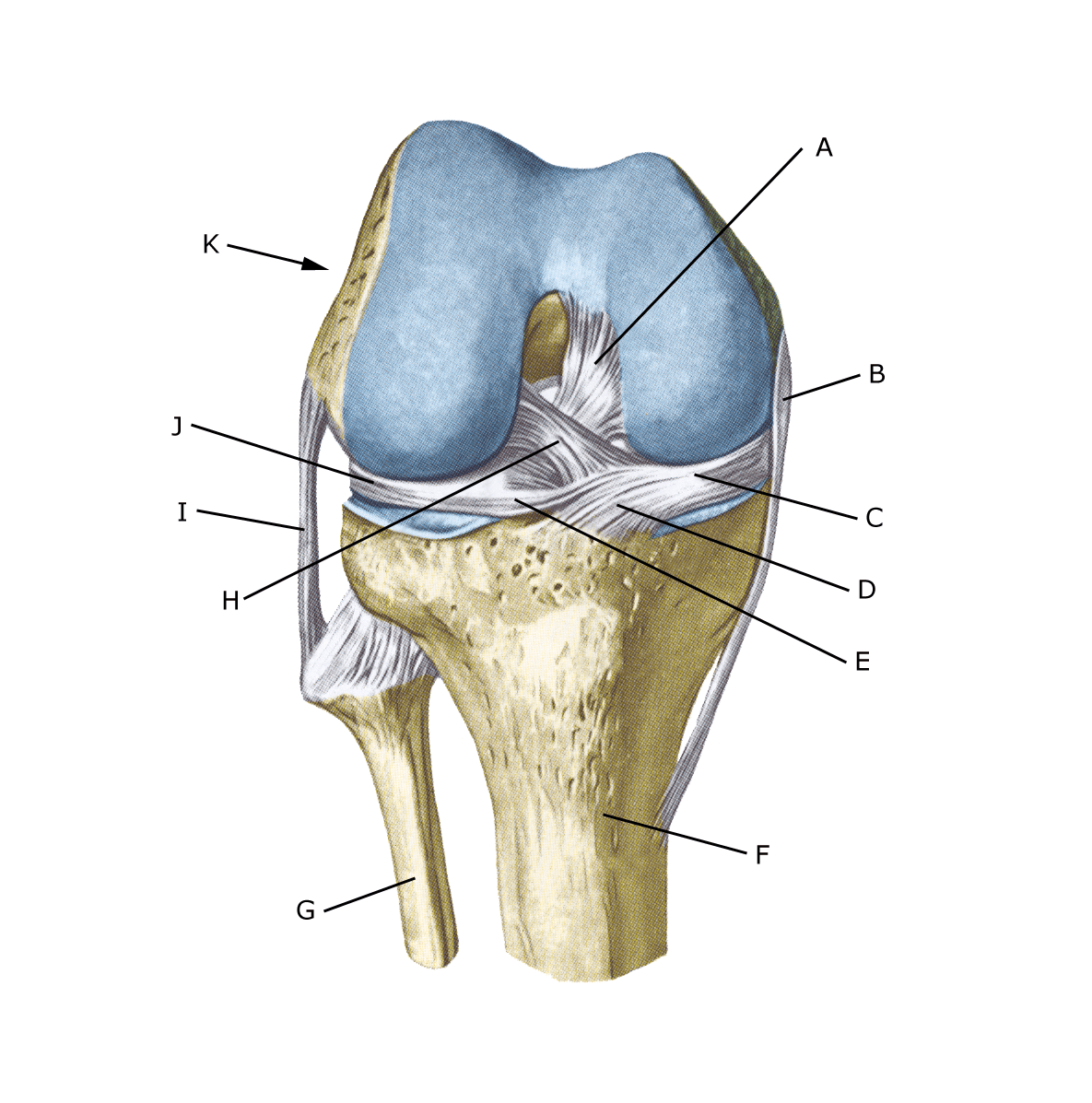
Knee joint from the front

A. Ligamentum cruciatum posterius
B. Ligamentum collaterale mediale/tibiale
C. Meniscus medialis
D. Insertio anterior menisci medialis
E. Ligamentum transversum genus
F. Tibiae
G. Fibulae
H. Ligamentum cruciatum anterius
I. Ligamentum collaterale laterale/fibulare
J. Meniscus lateralis
K. Femur

Cause

ACL tears are usually caused by a punch or kick directly into the front of the tibia just below the knee, pushing the tibia backwards in relation to the femur – see photo

Symptoms

Usually a pop is heard or felt and further sports activity must be cancelled. The knee may swell within the first few hours, after which the knee cannot fully bend. Relatively often there is no swelling in the knee.

Afterwards, you can often feel the leg giving way (knee failure). The prognosis is good and 90% can resume sports at the same level.

Examination

If you suspect a total or partial rupture of the ACL, you should see a doctor (or hospital) for a diagnosis. The doctor can perform various knee tests (posterior drawer laxity) to examine the stability of the knee.

It should be noted that looseness in the knee can often only be detected after 2 weeks. The examination is often more difficult in children. Ultrasound scanning can in many cases detect the rupture of the posterior cruciate ligament. See ultrasound scan. However, an MRI scan is usually necessary.

Treatment

Treatment of ACL tears primarily involves offloading and rehabilitation. In cases of severe looseness and symptoms or if the tear is combined with other knee injuries, surgery may be considered (Rupp GE, et al. 2024).

Bandage

The hinge bandage (Don-Joy) can be used for the first few weeks. Tape treatment of ACL ruptures in the knee has no certain effect.

Complications

In case of lack of progress, you need to consider

Especially

As there is a (modest) risk of permanent injury, the injury should be reported to your insurance company.

Rehabilitation

Rehabilitation program