Anatomy
The bones of the foot include 7 tarsal bones (ossa tarsi), 5 metatarsal bones (ossa metatarsi) and the bones of the toes (phalanx).
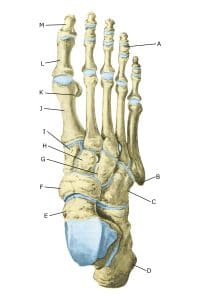
The foot, from the top.
A. Phalanx media
B. Tuberositas ossis metatarsalis V
C. Os cuboideum
D. Calcaneus
E. Talus
F. Os naviculare
G. Os cuneiforme laterale
H. Os cuneiforme intermedium
I. Os cuneiforme mediale
J. Os metatarsalei
K. Os sesamoideum
L. Phalanx proximalis
M. Phalanx distalis
Cause
For unknown reasons, the blood supply to a bone is reduced and it collapses (aseptic bone necrosis). Overloading may be a factor.
Most commonly, the collapse occurs in the navicular bone (Köhler I disease) or in the metatarsal bones (usually numbers 2 and 3) at the toe joints (Freiberg/Köhler II disease).
Symptoms
Pain in the midfoot (Köhler) or forefoot (Freiberg) on pressure (direct and indirect tenderness) and loading (walking, running). Mb Köhler most commonly affects boys aged 4-7 years and is 50% bilateral, while Mb Freiberg most commonly affects girls in their teens and is only 10% bilateral.
Examination
If localised foot pain increases or the bone surface is irregular on ultrasound, a medical examination is recommended to determine diagnosis and treatment. An X-ray and possibly an MRI scan will often be necessary.
Treatment
Treatment is primarily relief from pain-inducing activity. In some cases, supportive insoles and bandaging may be used. Usually, symptoms subside after 6-9 months and bones can normalise after ½-4 years. Surgery is therefore relatively rarely indicated (Yoshimura I, et al. 2024).
Rehabilitation, specific:
Of course, it’s important to keep all other muscles and joints working (Rehabilitation, general) while the collapsed bones are stabilised and restored.
You can keep fit by cycling and swimming.
Complications
If progress is not smooth, you should be reassessed by your doctor to ensure that relief is still sufficient