Fracture of the vertebral arch

Anatomy

The back is made up of box-shaped vertebrae that are held in place partly by the shape of the bones, partly by ligaments and partly by the large and small back and abdominal muscles.

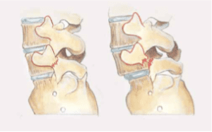
Fracture of the vertebral arch (archolysis) of the 5th lumbar vertebra. Incipient forward slippage (spondylolisthesis) of the vertebral body (right).

Fracture of the vertebral arch (arkolysis) on both sides (top view).

Pictures from Idrætsskadesbogen, FADLs Forlag.

Cause

There are different causes of lumbar vertebrae loosening: congenital (dysplastic), acute fractures or more frequent fatigue fractures (stress fracture) often due to congenital weakness of the vertebral arch (spondylolysis/archolysis). These types of fractures are mainly seen in children and adolescents, while wear-related looseness is mainly seen after the age of 50. In rare cases, fracture-induced looseness can be caused by diseases of the vertebrae.

Vertebral arch fractures are the most common cause of lumbar vertebrae looseness among young people and athletes who repeatedly bend their backs backwards (gymnastics, throwing, dancing, high jump, football, weightlifting, rowing).

In some cases, the looseness can cause one of the vertebrae to slip forwards (spondylolisthesis) and although it is frequently asymptomatic (seen in up to 5-10% of the population and in 35% of Inuit), it can trigger back symptoms and in severe cases pressure on nerves in the back (Li N, et al. 2022).

Symptoms

Average age at the time of diagnosis is approximately 15 years. Pain and stiffness in the lower back (‘lumbago’) radiating to the buttock muscles and thighs. The pain worsens during the day. The pain is relieved by forward bending (flexion), while backward bending (extension) is impaired (cancelled) and triggers the known pain.

Mobility is often almost eliminated in extension and reduced in lateral flexion. Nerve pressure can radiate to the legs (sciatica) and in rare cases urinary incontinence (cauda equina).

Examination

Mild back pain does not necessarily require a medical examination, but all cases of persistent, repetitive or severe back pain should be investigated. The doctor will be able to determine if there is an indication for further examination, such as X-rays (including oblique scans), which can establish the diagnosis and rule out other causes (e.g. Paget’s disease). X-rays are rarely normal, which is why MRI or CT scans must be supplemented (Mohile NV, et al. 2022)

Treatment

Treatment is dependent on the severity. However, the vast majority of cases can be treated with (rapid) relief (often for a few months) from pain-inducing activity and gentle strengthening of the abdominal muscles (which in severe cases may take several months).

In some cases a brace can be used and in about 10% of cases where the forward slippage (spondylolisthesis) is significant and symptoms do not resolve with prolonged offloading or neurological symptoms, surgery may be necessary. (Choi JH, et al. 2022).

Complications

If the pain does not subside after relief and treatment, you should be (re)examined by a doctor.

In particular, the following should be considered:

However, there are many other causes of lower back pain (arthritis, infections, metabolic disorders, nerve disorders, tumours), some of which will require further specialist investigation.

Rehabilitation

Rehabilitation program