Anatomy
The lower leg is made up of many muscles, but especially the two largest calf muscles (Gastrocnemicus and Soleus muscles), which together form the Achilles tendon that attaches to the back of the heel bone (calcaneus), are relatively prone to injuries in the form of tears and/or haematomas in the muscles.
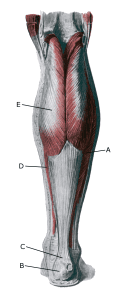 Lower leg from behind:
A. M. soleus
B. Tuber calcanei
C. Tendo calcaneus (Achillis)D. M. gastrocnemius
Cause
When a muscle is subjected to a force that exceeds its strength, a tear occurs, often accompanied by bleeding. The vast majority of muscle strains are partial muscle strains.
Injuries in muscles can be localised in the muscle tissue itself, in the fascia around/between the muscles or in the tendon part of the muscles, which has a major impact on prognosis (muscles heal much faster than tendons).
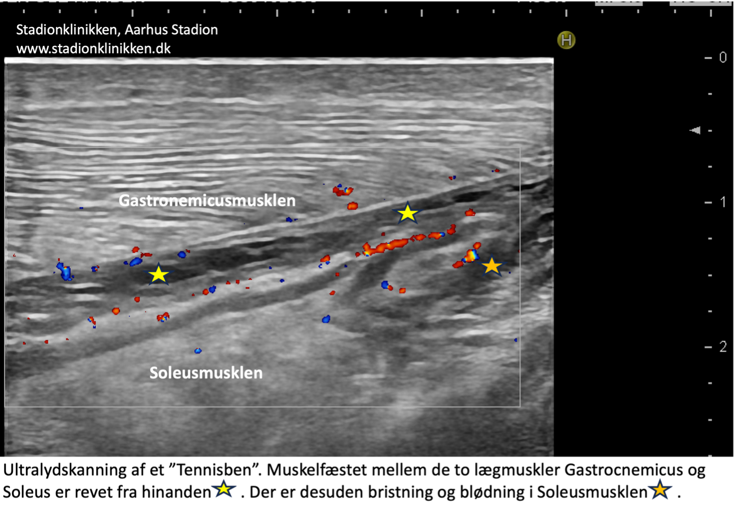
One of the weakest points is the attachment between the inner (medial) part of the calf muscle (Gastrocnemicus) and the underlying Soleus muscle (where the injury is called a ‘Tennis bone’) (Dai M, et al. 2021).
Muscle haemorrhage can also occur due to muscle damage after direct trauma to the calf muscle (e.g. kicking). In some cases, the haemorrhage can attract fluid so that the accumulation grows in the following days (hygroma) in the fascia between the two muscles.
Symptoms
For muscle injuries without direct trauma to the muscle, in mild cases a localised soreness will be felt after the strain (‘muscle strain’, ‘threatening fibre’). In more severe cases, you will feel a sudden, shooting pain in the muscle (‘partial muscle rupture’, ‘fibre rupture’) and in the worst case, you will feel a violent snap, after which it is impossible to use the muscle (‘total muscle rupture’).
In muscle injuries, the following three symptoms are characteristic: Pain when pressing, stretching and activating the calf muscle against resistance (pushing off, toe walking). With total tears, you can often see and feel a defect in the muscle and swelling due to the contracted muscle bulge and bleeding.
When the inner (medial) attachment between the two large calf muscles (Tennis elbow) tears, there will be (often long-lasting) pain inside the calf.
With direct trauma to the calf muscle, there will be pain corresponding to the trauma and there will often be a large blood pool.
Examination
Usually, the diagnosis is made on clinical examination alone, where there is characteristic muscle pain on pressure, stretching and activation of the muscle. If there is any doubt about the diagnosis, an ultrasound or MRI scan can be used as a supplement.
See ultrasoundscan of a “tennisben”
Treatment
Treatment for calf muscle injuries includes relief from pain-inducing activity, stretching and slowly increasing rehabilitation within the pain threshold.
Localising whether a tear is in the muscle, in the fascia or in the muscle tendon is of great importance for the speed of rehabilitation. This can often be determined by clinical examination. If in doubt, ultrasound or MRI scans can pinpoint the location of the injury.
Large blood clots (or hygromas) can be ultrasound-guided drained, reducing pain and shortening the injury period. Medical treatment and surgery are usually not indicated.
Tennis elbow can take months to rehabilitate before maximum strain is possible again.
Complications
If the progression is not smooth, you should consider whether the diagnosis is correct
In particular, the following should be considered:
- Complications of muscle rupture
- Rupture of the achilles tendon
- Deep vein inflammation.