Meniscus lesion

Anatomy

The bones of the knee joint include the femur (thigh bone), tibia (shin bone) and patella (kneecap). The articular surfaces of the femur, tibia and patella are covered with a few millimetres of cartilage that serves to reduce stress on the articular surfaces. Both inside and outside the joint there is an annular cartilage disc (meniscus). The knee joint is reinforced by a joint capsule that is laterally reinforced with an external and internal collateral ligament (ligamentum collaterale laterale/fibulare and mediale/tibiale). The inner meniscus attaches to the inner collateral ligament, but the outer meniscus does not attach to the outer collateral ligament.

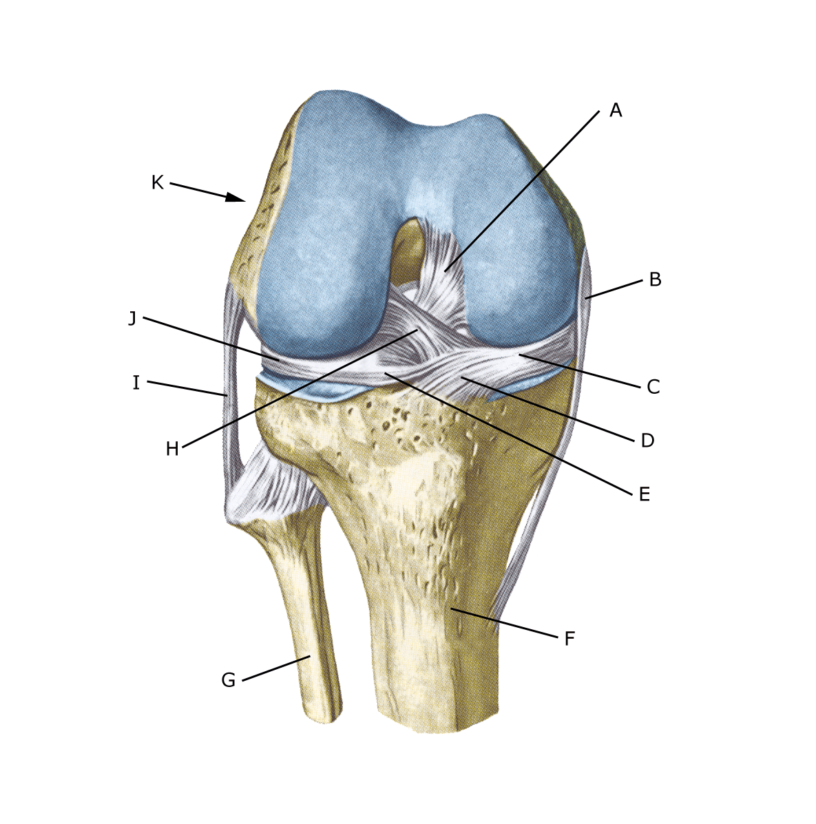
Knee joint from the front:

A. Ligamentum cruciatum posterius
B. Ligamentum collaterale mediale/tibiale
C. Meniscus medialis
D. Insertio anterior menisci medialis
E. Ligamentum transversum genus
F. Tibiae
G. Fibulae
H. Ligamentum cruciatum anterius
I. Ligamentum collaterale laterale/fibulare
J. Meniscus lateralis
K. Femur

Knee joints from above:

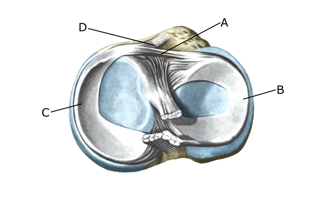
A. Ligamentum transversum genus
B. Meniscus lateralis
C. Meniscus medialis
D. Insertio anterior menisci

Cause

Meniscus tears usually occur acutely when the knee is stretched while rotating. This squeezes the meniscus between the femur and tibia. Internal meniscus injuries are far more common than external ones. Meniscus injuries in children are rare. In children with congenital changes in the meniscus (discoid meniscus), injuries can occur with minor stresses (Geffroy L. et al. 2021).

Symptoms

Pain at the joint line when pressing and rotating the knee. Aggravated in a squatting position and when climbing stairs. The pain often occurs suddenly when twisting the knee while stretching or bending and may be accompanied by swelling in the knee. In some cases, the knee may lock (because a flap of the meniscus becomes trapped).

Often there are a few degrees of stretch defect, the cause of which should be determined as soon as possible (within 2 weeks) for treatment. treatment. In other cases, you may feel a small, tender swelling at the joint line (meniscus cyst). With long-term discomfort, the thigh muscle will often shrink.

Examination

If injuries inside the knee joint are suspected, a medical examination is always necessary and tenderness at the joint line and bending or stretching defects in the knee are often found. Often general clinical examination is not sufficient.

A knee X-ray should be taken in two planes after acute knee injury (usually within 1 week) if any of the following apply: the patient is under 12 years old, cannot walk 4 steps due to pain, pressure tenderness of the patella or fibular head (caput fibulae), the knee cannot bend more than 60°, or there is ligament damage (Pittsburgh knee rules).

Often general clinical examination is not sufficient to diagnose a meniscus tear, which is why an MRI or ultrasound scan is required. See ultrasound scan.

Treatment

Treatment includes relief from pain-inducing activity and rehabilitation of the knee including strength and balance training. If the discomfort does not slowly subside, if there are locking episodes or if the meniscus tear is large (e.g. assessed by MRI or ultrasound scan), surgery should be considered. Surgery will attempt to suture the torn part of the meniscus. Removing parts of the meniscus has become very reluctant as it increases the long-term risk of osteoarthritic changes in the knee (Sihvonen R, et al. 2020).

Rehabilitation

Strength training of the muscles around the knee, stretching and balance training. Exercise should not cause increasing swelling (or pain) in the knee. If surgery has been performed, rehabilitation should be discussed with the operating surgeon.

Complications

If no progress is made, you need to consider whether the diagnosis is correct.

In particular, the following should be considered:

If surgery has been performed, infection should be considered.

Especially

As there is a risk of permanent damage, the injury should be reported to your insurance company.

Rehabilitation

Rehabilitation program