Anatomy
The lower leg consists of two bones: the tibia and fibula. The calf muscles are divided into 3 groups (muscle compartments) of strong muscle membranes (fascia) that are partially rigid: an anterior muscle compartment (comprising the flexor muscles of the foot, the extensors), an external (lateral) muscle compartment (comprising the peroneal muscles) and a posterior muscle compartment (comprising the extensor muscles of the foot, the flexors), which is divided into a superficial and a deep part.) Each muscle group has its own blood and nerve supply.
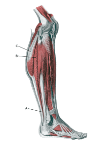
Lower leg:
A. Tendo calcaneus (Achillis)
B. M. soleus
C. M. gastrocnemius
Cause
During muscle training, muscles can grow so fast that the surrounding muscle membranes (fascia) cannot give way sufficiently. This increases the pressure in the muscle tissue, which can affect the muscles, blood vessels and nerves (chronic compartment syndrome). This condition is particularly common in intensively trained athletes.
In the event of acute severe strain or direct trauma (e.g. bone fractures), fluid accumulation and haemorrhaging in the muscle compartment can occur, causing significantly increased pressure in the muscle compartment with acute effects on muscles, nerves and blood vessels (acute compartment syndrome).
Symptoms
In chronic compartment syndrome, there is a slow onset of muscle pain after a few minutes of activity. You feel the muscle ‘tighten’ and become hard, which is accompanied by discomfort and possibly sensory disturbances. The pain subsides a few minutes after stopping the strain, but quickly returns if the strain is resumed.
Acute compartment syndrome is characterised by increasing pain that is often more severe than expected based on the primary assessment of the extent of the injury. Even stretching hurts. At the same time, the pain does not subside shortly after stopping exertion.
Examination
The diagnosis of chronic compartment syndrome is primarily based on the characteristic strain-related pain and a clinical examination by a relevant professional. Muscle compartment pressurisation may be performed, but pressure measurement is associated with significant limitations and lack of documentation (Ritchie ED, et al. 2023). Imaging (X-ray, ultrasound and possibly MRI scans) rarely provides a definitive answer.
If acute compartment syndrome is suspected, an urgent medical examination must be performed (possibly in hospital).
Treatment
The treatment for chronic compartment syndrome is relief from pain-inducing activity, stretching and slowly increasing strength training within the pain threshold to allow the muscle barriers to give way. If there is persistent discomfort or frequent relapses despite regular rehabilitation, one or more of the muscle splints can be divided (Weiss C, et al. 2022).
Acute compartment syndrome always requires urgent medical examination (possibly in a hospital) with the possibility of emergency surgical splitting of the muscle membranes, otherwise permanent damage to muscles and nerves may occur.
Rehabilitation
If surgery is performed for both acute or chronic compartment syndrome, there may be specific rehabilitation instructions from the surgeon depending on the type and course of surgery.
Complications
Acute compartment syndrome can cause permanent damage to muscles and nerves if not treated as soon as possible. Amputations have been described after acute compartment syndromes.
Especially
As there is a risk that acute compartment syndromes can cause permanent damage, the injury should be reported to your insurance company.