Anatomy
The hip joint consists of the hip socket (acetabulum) and the head of the femur (caput femoris). The joint surfaces are covered by a layer of cartilage a few millimetres thick, which reduces the stress on the joint surfaces.
Cause
For unknown reasons (e.g. hereditary conditions, fluid accumulation in the hip joint, passive smoking, (Gao H, et al. 2020)) there is a slow collapse (aseptic bone necrosis) of the femoral head, which collapses and flattens, causing irritation in the hip joint.
It affects physically active children aged 3-11 years. Boys are affected 3 times more often than girls. 15% are bilateral.
Symptoms
Initially, there is often only mild pain and fatigue in the hip joint with movement and strain, as well as a limp and very often limited rotation in the hip joint. Later, pain and limping increase due to shortening of the leg. Sometimes the pain is initially felt in the knee and thigh.
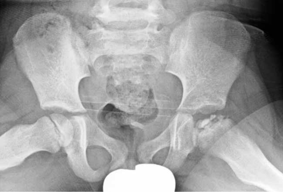
Examination
It is important to get a medical examination and diagnosis as soon as possible, as prompt treatment is crucial for a good outcome. It will often be necessary to supplement the regular clinical medical examination with an ultrasound or MRI scan, where previous bone changes and fluid in the hip joint are easily seen, and possibly an X-ray (see X-ray with RTG disease), where the late bone changes are seen.
Treatment
Treatment should start as soon as possible and is primary relief, which should start as soon as it is suspected. Calvé-Legg-Perthes disease requires highly specialised treatment at the orthopaedic surgery departments in Aalborg, Aarhus, Odense and Rigshospitalet. Treatment primarily involves intensive support, possibly in a wheelchair. Surgery may be indicated.
Rehabilitation
The amount of exercise and rehabilitation that can be allowed is entirely dependent on the severity of the disease, which is why all rehabilitation should be done in close co-operation with the doctors supervising the treatment.
Complications
In most cases, there will be no discomfort in the form of pain or activity limitations in everyday life. The younger the child is when the disease occurs, the better the prognosis. The condition carries a risk of shortening of the leg and osteoarthritis of the hip joint. However, the chance of the femoral head healing with a normal shape is very high, especially for the youngest children. If no progress is made, you need to consider whether the diagnosis is correct
In particular, the following should be considered:
- Bacterial infection of the joint (pyartron)
- Fluid accumulation in the joint
- Calve-Legg-Perthes disease
- Epifysiolysis caput femoris
- Inflammation of the muscle attachment at the growth zone of the sitting bone
- Inflammation of the growth zone around the hip
- Inguinal hernia
- Bone disease (Yagdiran A, et al. 2020).