Luxation of the knee cap

Anatomy

The large anterior thigh muscle (musculus quadriceps femoris) is made up of four muscles (vastus lateralis, vastus medialis, vastus intermedius and rectus femoris). The muscles all attach to the upper edge of the kneecap. The patella tendon (ligamentum patellae) connects the lower edge of the patella to the upper, anterior part of the tibia (tuberositas tibiae). The patella is held in place by the structures that attach to the patella, specifically the anterior thigh muscle, the joint capsule and several ligaments (retiaculum patellae mediale and retiaculum patellae laterale) and the patella tendon (ligamentum patella).

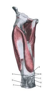
Knee from the front:

A. Tendo m. adductoris magni
B. Retinaculum patellae mediale
C. Meniscus medialis (Inner meniscus)
D. Ligamentum collaterale mediale/tibiale (Medial collateral ligament)
E. Bursa anserina
F. Bursa subtendinea m. sartori
G. Ligamentum patellae
H. Patella (Kneecap)

Cause of the problem

Kneecap dislocation can occur after a blow to the knee, but more commonly after a sudden forceful knee movement where the knee is simultaneously twisted and stretched. The kneecap can be displaced to the outside (laterally) of the knee, causing the ligaments holding the kneecap to rupture. Often the kneecap hits the femur, resulting in a risk of cartilage damage in the knee: Cartilage damage in the knee.

Symptoms

Sudden onset of severe pain that makes continued sports activity impossible. In some cases, the kneecap may dislocate completely on the outside of the knee, locking the knee in a bent position (total joint dislocation) until the kneecap suddenly slips back into place and the knee can be stretched again. In other cases, the patella is only partially displaced on the outside of the knee and spontaneously moves back into place when the knee is extended (partial dislocation, subluxatio patellae).

Examination

If the kneecap is in place, diagnosis can be difficult and anyone with a suspected dislocated kneecap should always be medically examined to ensure diagnosis and proper treatment. During the examination, severe pain will typically be triggered when the patella is pressed outwards (laterally) while bending the knee (Apprehension test).

The kneecap will often extend further outwards on the injured knee than on the healthy knee. If a kneecap dislocation is suspected, an X-ray should be performed to detect any bone spurs, which 30% will have. The X-ray can also identify anatomical abnormalities. MRI scans can provide information about cartilage conditions in the knee after total and partial kneecap dislocations

Treatment

Partial joint slips should primarily be treated with offloading and rehabilitation.

In the case of a total joint dislocation, the kneecap must be replaced immediately if it has not happened spontaneously. First-time total knee replacements can be rehabilitated, but about 1/3 relapse. Often a bandage is applied for a few weeks during which the knee’s mobility is restricted.

In case of repeated (total or partial) joint dislocations, surgery is advised (Aicale R, Maffulli N. et al. 2020). After acute traumatic dislocation, the athlete can start sport-specific training after surgery after 1-2 months and in most cases resume sport after 3-6 months.

Bandage

Tapes and bandages have no proven preventive effect in cases of previous total or partial kneecap dislocation, but are still widely used according to experience, See tape.

Especially

As there is a risk of permanent injury, the injury should be reported to the insurance company.

Rehabilitation

Rehabilitation program