Anatomy
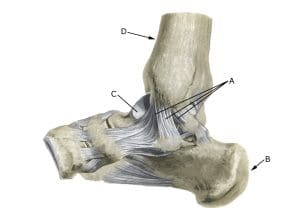
A. Ligamentum mediale/deltoideum
B. Calcaneus
C. Talus
D. Tibia
Cause
A tear in the lateral ligaments of the ankle joint is one of the most common sports injuries and occurs when the foot rolls outwards (supination), causing the lateral ligaments to be overstretched and tear. In mild cases, this is referred to as a sprain or strain, and in severe cases as a complete or partial tear.
The anterior ligament (anterior talofibular ligament) ruptures most frequently. The middle ligament (calcaneofibular ligament) ruptures in 20% of cases where the anterior ligament ruptures. The posterior ligament usually only ruptures in severe ankle injuries with fractures or dislocations.
Consequential injuries associated with ligament ruptures include rupture of the ligaments holding the tibia and fibula together (syndesmosis rupture), fluid accumulation in the joint (traumatic synovitis) and tendonitis, midfoot ligament rupture, ankle fractures, cartilage damage inside the ankle joint (major cartilage lesions are seen in 7% of sprains) and damage to the subtalar joint (the joint between the heel bone (calcaneus) and ankle roll bone (talus)).
In some cases, ankle ligament ruptures are complicated by inflammation of the joint capsule around the ankle joint (capsulitis).
Symptoms
Sudden onset of pain on and below the outer ankle bone (malleolus lateralis), swelling and discolouration due to bleeding and pain when walking.
Examination
Diagnosis is made on clinical examination, with mild cases (sprains) showing minimal swelling and no discomfort with normal walking, but usually pressure tenderness at the anterior cruciate ligament attachment (ATFL). The amount of swelling is not a measure of the extent of the injury. Pronounced swelling or pain should be examined by an appropriate professional to rule out, among other things, ankle fractures, especially fractures in the growth zone of the lower leg at the ankle and rupture of the ligaments between the tibia and fibula (syndesmosis rupture).
If a fracture is suspected, an X-ray of the ankle joint should be taken. Previous vigorous twisting of the ankle joint in the acute stage to assess the degree of looseness is no longer indicated as this does not affect the choice of treatment.
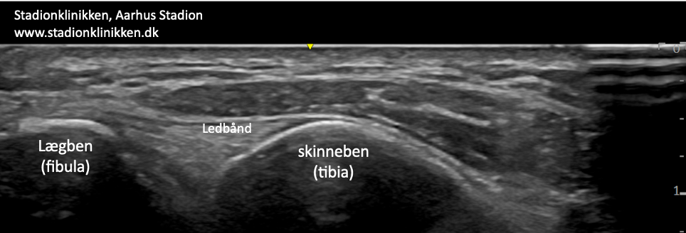
Ultrasound scanning can visualise external ligaments and the ligament (lig. tibiofibulare ant. inf.) between tibia and fibula (syndesmosis) and inflammation in capsulitis (Colò G, et al. 2023). With dynamic ultrasound scanning, looseness of the ligaments can be assessed (the ligament is stressed during simultaneous ultrasound scanning, whereby it can be seen whether the joint gap increases as a sign of ligament rupture, see video (Heitz PH, et al. 2024).
Treatment
The treatment of ligament injuries is now conservative (rehabilitation). In the past, many patients underwent surgery and were bandaged, an approach that has largely been abandoned for uncomplicated ligament tears. Surgery is often indicated in cases of recurrent ankle sprains and syndesmotic tears with significant laxity (Colò G, et al. 2023).
Bandage
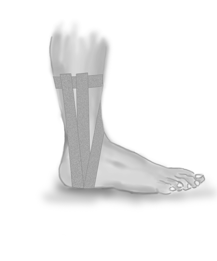
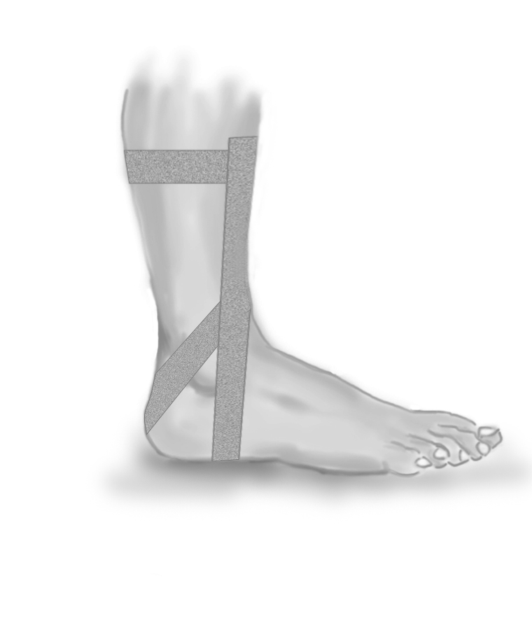
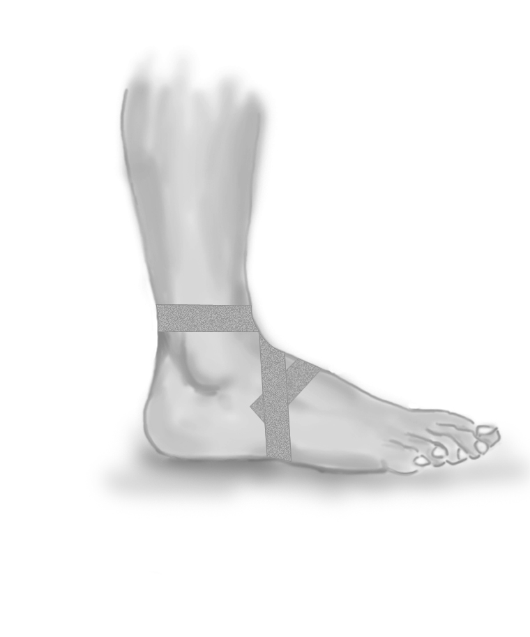
During the rehabilitation programme, tape (or various types of ankle supports) is often used when starting to run on uneven surfaces or when running with rapid changes of direction. The ligaments contain nerve cells (proprioceptors) that send information to the brain about the position of the ankle joint. Information is sent from the brain to the muscles, which are activated so that the ankle joint is held correctly.
When the ligaments are damaged, these nerve pathways don’t function optimally and the risk of renewed twisting of the foot increases. The primary function of the tape is to stimulate the small nerve cells (proprioreceptors) in the skin so that they can ‘stand in’ for the temporarily damaged nerve cells in the ligaments. The function of the tape is thus not just a purely mechanical ‘stabilisation’ of the ankle joint, see tape 1, tape 2 og tape 3.
In some cases, certain types of bandages around the ankle joint can be beneficial for 5-6 weeks. Specialised bandages have been shown in some studies to reduce the risk of ligament re-injury (Zwipp H. 2023).
Complications
If the progress is not smooth, you should consider whether the diagnosis is correct or if there are complications:
In particular, the following should be considered:
- Rupture of ligaments between the tibia and fibula (syndesmosis rupture),
- Bone fracture in the ankle,
- Bone sheath tearing (avulsion),
- Tendon rupture (peroneus luxation),
- Tendonitis,
- Fluid accumulation in the joint (traumatic synovitis),
- Joint cartilage damage, osteoarthritis (osteochondral lesion)
10-15% of athletes with injuries to the external ligaments of the ankle continue to have discomfort 1 year later, probably due to insufficient rehabilitation before resuming full sports activity.
In particular
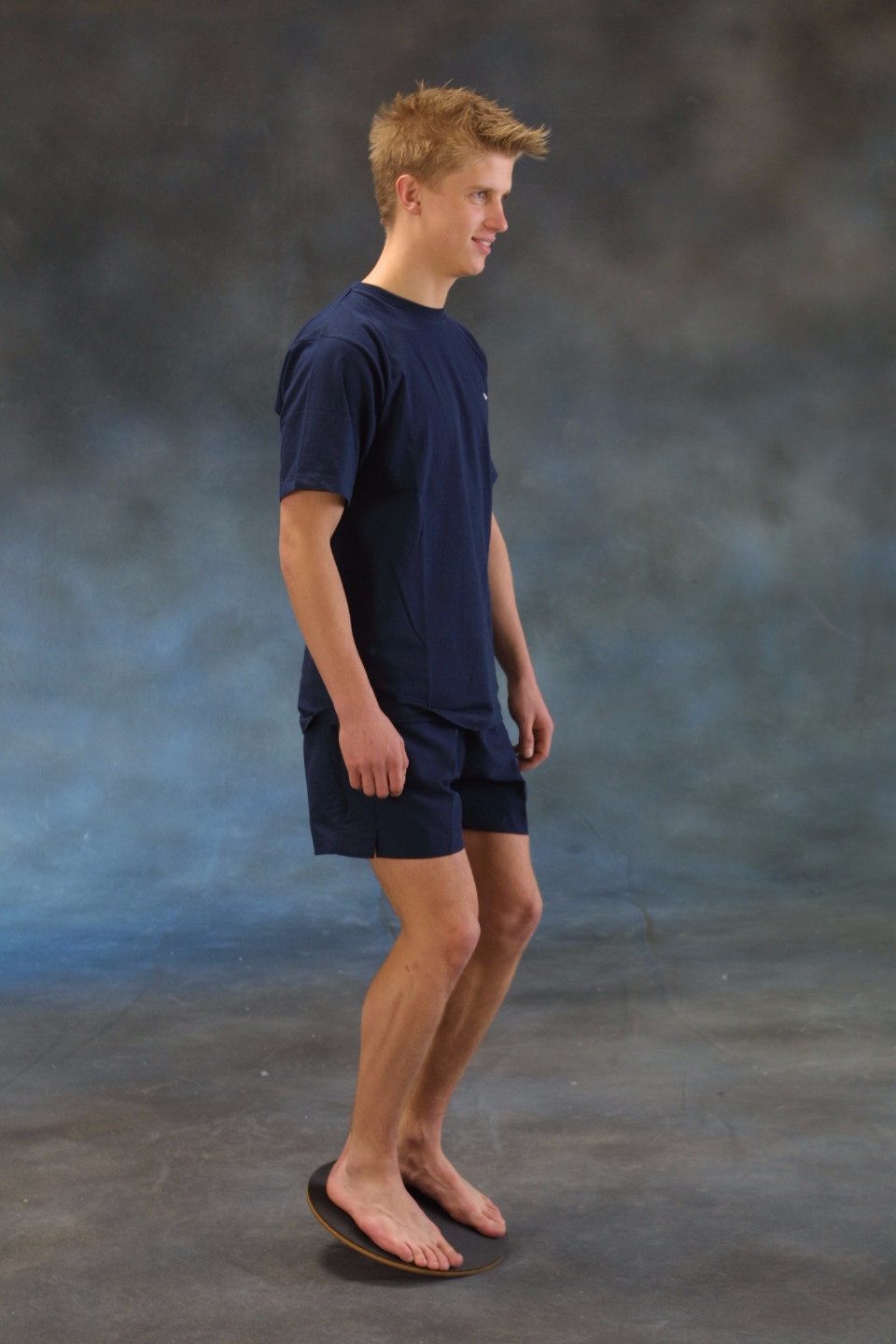
Balance board exercises 1 and 2 are important for both rehabilitation and prevention (Al Attar WSA, et al 2024). Balance board exercises should be carried out regularly for the remainder of an athlete’s active career as a preventive measure if there has been a history of ligament injuries in the ankle joint. Start by standing on both legs on the seesaw and support with your hands on the wall.
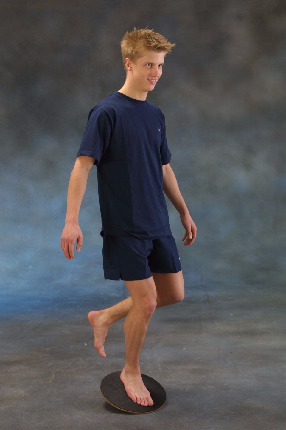
Gradually release the support with your hands and eventually train with support on one leg only. You can brush your teeth morning and evening on the rocker board (Wang F, et al. 2023).