Discus triangularis

Anatomy

The bones of the wrist consist of 8 carpal bones (ossa carpi), which together with the two forearm bones, the ulna and radius, form the wrist. On the little finger side (ulnar side) of the wrist, there is a triangular cartilage disc/meniscus (discus triangularis) a few mm in size between the ulna and the lunate, which together with various ligaments stabilises the wrist under load. The meniscus and ligaments have many names, including Triangular Cartilage Complex (TBK) and internationally Triangular Fibrocartilage Complex (TFCC) (van der Post AS, et al 2022).

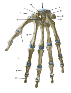
The bones of the right hand palm:

A. Os lunatum
B. Os triquetrum
C. Os pisiforme
D. Os hamatum
E. Phalanx distalis
F. Phalanx media
G. Phalanx proximalis
H. Os metacarpale II
I. Ossa sesamoidea J. Os trapezoideum K. Os trapezium
L. Os capitatum
M. Os scaphoideum
N. Carpus

Cause of the problem

Falling on an extended arm with the hand bent backwards (extension) and rotation such as handstand gymnastics, push-ups and climbing can acutely damage the ligaments and meniscus in the wrist.

The meniscus (discus triangularis) can also be worn by repetitive strain between the ulna and the carpal bones, causing gradual thinning and perforation of the meniscus (discus triangularis) while destroying the cartilage of the carpal lunate and the ligaments between the carpal lunate bones and the triquetrum ruptures. This causes the joint between the bones to become unstable, which can trigger osteoarthritic changes in the little finger side (ulnar side) of the wrist.

Symptoms

Pain in the little finger side of the wrist. The pain worsens when the wrist is used and the discomfort improves at rest. Later, the pain is constantly present with daily activity. There may be a clicking sensation in the wrist when moving.

With severe damage to the ligaments around the meniscus, the joint between the two bones of the forearm can be unstable, where twisting movements of the wrist provoke pain and the ulna prominently forms a bulge on the upper side of the wrist.

Examination

The diagnosis is usually made by a clinical examination where tenderness is found in the joint between the ulna and the lunate bone. Ultrasound and MRI scans can be used in addition, but often the diagnosis is first made by arthroscopic surgery (arthroscopy), which is best for assessing the ligaments between the bones.

Treatment

The vast majority of acute injuries to the meniscus and ligaments in the wrist can be treated with relief from pain-inducing activity for 4-6 weeks, possibly with a wrist brace. Long-term strain pain can be reduced by medical treatment in the form of NSAIDs or ultrasound-guided injection of adrenal cortex hormone around the damaged meniscus.

If offloading and medical treatment are unsuccessful, binocular surgery can be attempted to remove or repair the damaged meniscus (Schachinger F, Farr S. 2021).

Rehabilitation

Usually, you can continue cardio training in the form of cycling and running, as well as rehabilitation according to the guidelines under rehabilitation in general. Gentle wrist range of motion exercises ensure mobility is maintained and are combined with slowly increasing the load on the muscles around the wrist within the pain threshold.

Complications

If no progress is made, consider whether the diagnosis is correct, e.g. fracture of the bones around the wrist, tendonitis of the extensor carpi ulnaris (in the 6th joint on the back of the hand).

The bones of the wrist, where the forearm bone (ulna) is often compressed against the carpal bone (lunate) due to damage to the ligaments, damaging the meniscus (discus triangularis) (From the book Idrætsskader, 2nd edition, FADL’s forlag)

Rehabilitation