Anatomy
The articular surfaces are covered with a few millimetres of cartilage that serves to reduce stress on the articular surfaces.
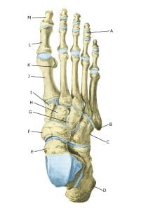
The foot, from the top.
A. Phalanx media
B. Tuberositas ossis metatarsalis V
C. Os cuboideum
D. Calcaneus
E. Talus
F. Os naviculare
G. Os cuneiforme laterale
H. Os cuneiforme intermedium
I. Os cuneiforme mediale
J. Os metatarsalei
K. Os sesamoideum
L. Phalanx proximalis
M. Phalanx distalis
Cause
Localised articular cartilage injuries are common in sports and can occur when the joint is twisted forcefully, causing the articular surfaces to collide and destroy the cartilage. Most articular cartilage injuries are localised in the ankle joint and often on the talus, but can occur in any joint on the foot. In ankle sprains, minor cartilage lesions are seen in about 50% and major cartilage damage in about 7%.
In some cases, a piece of cartilage can be torn off that can move around the joint (joint mouse) and become trapped (locking). In other cases, a piece of bone in the joint (osteochondritis dissicans) becomes dislodged without a known cause (repetitive microtrauma, ischaemia, subsequent avascular necrosis, genetic predisposition, endocrine or metabolic factors including vitamin D deficiency) (Porta-Alesandria J, et al., 2021).
Symptoms
Pain in the joint when moving with strain. Inflammation (synovitis) of the synovial membrane may also occur, leading to fluid build-up in the joint. Loose pieces of cartilage can cause the joints to lock up.
Examination
General clinical examination is often not enough. Imaging (primarily X-rays and MRI and possibly CT scans) is therefore necessary to make a diagnosis. In some cases, arthroscopy (binocular examination) primarily of the ankle joint is indicated.
Treatment
For trauma-related cartilage damage, treatment includes relief from pain-inducing activities until pain-free. After this, gentle rehabilitation can begin. There is no treatment that can restore the damaged cartilage, which has little ability to heal.
Surgery is usually only considered if there is no progress after at least 3-6 months of rehabilitation or if cartilage and bone fragments are torn. In general, the effect of surgical treatment is only sparsely documented (Steele JR, et al. 2023).
If a piece of bone in the joint (most commonly the ankle joint) loosens without prior trauma (osteochondritis dissicans), it is important that the diagnosis is made as soon as possible, as early treatment improves the outcome. Treatment may include bracing or surgery, depending on the extent of the lesion and whether a piece of both cartilage and bone has become loose (Steele JR, et al. 2023).
Rehabilitation, specific:
The amount of weight-bearing and rehabilitation that can be allowed depends entirely on the severity, location and treatment of the cartilage lesion, so all rehabilitation should be carried out in close co-operation with the doctors supervising the treatment. All training that does not involve loading the foot can continue unchanged.
Complications
Large cartilage injuries located on the weight-bearing parts of the joint are one of the most serious sports injuries and often lead to sports retirement. If there is no progress on treatment, various differential diagnoses should be considered such as bone fractures, congenital bone deformities, rupture of the syndesmosis, looseness of the lateral collateral ligaments of the ankle, tendinopathy of the peroneal tendons behind the outer ankle bone, fluid in the foot or metatarsal joint.
In particular
As there is a risk that the injury may cause permanent damage, you should report the injury to your insurance company.