Anatomy
The tendons around the ankle are attached to the bones in several places with small connective tissue layers (retinacles). Behind the outer ankle bone are the two peroneal muscles (peroneus longus and peroneus brevis), which help stabilise and bend the ankle.
Cause of the problem
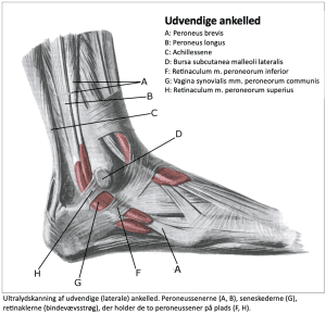
If there is a rupture of the connective tissue strands (retinaculum musculorum peroneorum superius and inferius) behind the lateral malleolus, the muscle tendons (peroneus brevis and peroneus longus) can slip over the lateral malleolus, causing discomfort. Rupture of the connective tissue layer is relatively often combined with outer ligament ligament injury in the ankle joint.
Symptoms
Pain at the lateral malleolus, where certain movements of the ankle can cause a grinding, painful sensation on the lateral malleolus.
Examination
When the painful movement of the ankle is performed, the tendon can often be seen slipping over the outer ankle bone on general examination. The diagnosis is usually made during a general medical examination.
If there is any doubt about the diagnosis, an ultrasound scan can be performed while moving the ankle (dynamic ultrasound scan). The ultrasound scan will show if there is damage to the tendon or if there is bleeding in the acute stage. Tendonitis can develop later in the process.
Treatment
Treatment primarily involves relief from pain-inducing activities (running). Some recommend bandaging for 6 weeks (Bakker D, et al. 2019). If there is no steady progress on offloading and rehabilitation and an ultrasound scan shows tendonitis, treatment can be supplemented with medical treatment in the form of arthritis pills (NSAID) or injection of adrenal cortex hormone into the tendon sheath.
In some cases, surgical treatment can be attempted if offloading, rehabilitation and medical treatment do not lead to the desired result (Lootsma J, et al. 2023).
Complications
If no progress is made, consider whether the diagnosis is correct or if complications have arisen:
In particular, the following should be considered:
Supplement with additional examinations (X-ray, ultrasound and MRI scans)