Anatomy
The hip joint consists of the hip socket (acetabulum) and the head of the femur (caput femoris). The joint surfaces are covered by a layer of cartilage a few millimetres thick, which reduces the stress on the joint surfaces. The joint capsule is lined on the inside by a synovial membrane (synovium), which produces fluid to ‘lubricate’ the joint and nourish the articular cartilage
Cause
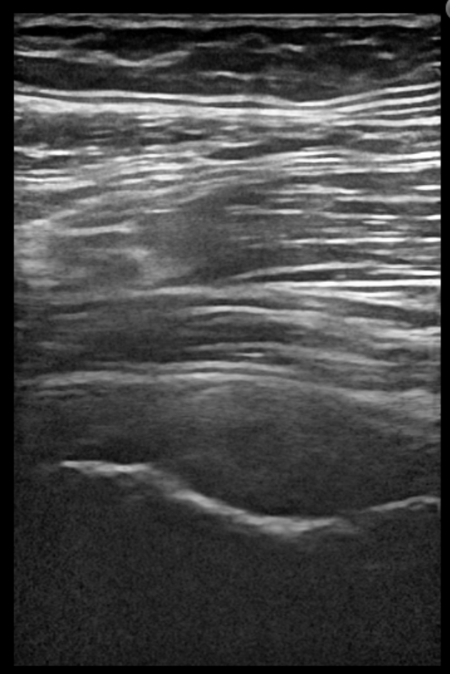
For partly unknown reasons, the lining of the hip joint produces too much fluid (coxitis). 50% have had an upper respiratory tract infection shortly beforehand (immunological cause), many minor repetitive stresses or one severe stress (traumatic cause) and an allergic reaction are some of the possible causes that trigger inflammation of the lining (synovitis), fluid formation (coxitis), restriction of movement and pain in the hip joint see ultrasound scan.
The condition is relatively common in children up to the age of 8, and boys are affected twice as often as girls.
Symptoms
Smerter i leddet ved bevægelse med belastning. Ofte vil der være bevægelsesindskrænkning ved rotation i hofteleddet. The pain is usually insidious, but can be acute. There may be a slight increase in temperature (38 degrees), but general condition is not affected.
In 5% of cases, the condition is bilateral. Usually, the symptoms resolve within 2-6 weeks with no ill effects. Approximately 10% may later experience a recurrence of coxitis (which still has a good prognosis).
Examination
It will often be necessary to supplement the regular clinical examination with an ultrasound scan, where the fluid in the hip joint is easily visible. The examination should rule out infection (possibly elevated temperature, affected general condition, severe pain), epiphysiolysis, Calvé-Legg-Perthes fracture and bone disease.
Treatment
Treatment primarily involves relief from pain-inducing activities until the swelling in the joint has subsided. Other muscles and joints are kept active. Once the pain has subsided, increasing load within the pain threshold can be initiated.
If relief is not successful or pain is severe, treatment can be supplemented with medication in the form of paracetamol and anti-rheumatic drugs (NSAIDs) or ultrasound-guided joint fluid drainage (which can be checked for infection and arthritis), which is rarely necessary.
Rehabilitation
Once the pain subsides, walking and then running can be gently resumed within the pain threshold according to General rehabilitation of children.
Complications
If no progress is made, you need to consider whether the diagnosis is correct.
In particular, the following should be considered:
- Bacterial infection of the joint (pyartron)
- Fluid accumulation in the joint
- Calve-Legg-Perthes disease
- Inflammation of the muscle attachment at the growth zone of the sitting bone
- Inguinal hernia
- Bone disease (Yagdiran A, et al. 2020).