Rupture of the Achilles tendon

Anatomy

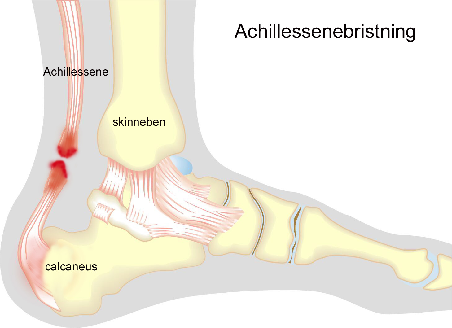
The calf muscle (M. Gastrocnemicus) consists of two muscle heads that come together in a broad tendon band that continues into the Achilles tendon. Another large calf muscle (M. Soleus) attaches to the front edge of the Achilles tendon and forms part of the Achilles tendon. The Achilles tendon thus consists of the 3 calf muscles (Triceps Surae). The Achilles tendon attaches to the heel bone (calcaneus). The weakest point of the Achilles tendon is about 3 cm above the attachment to the calcaneus.
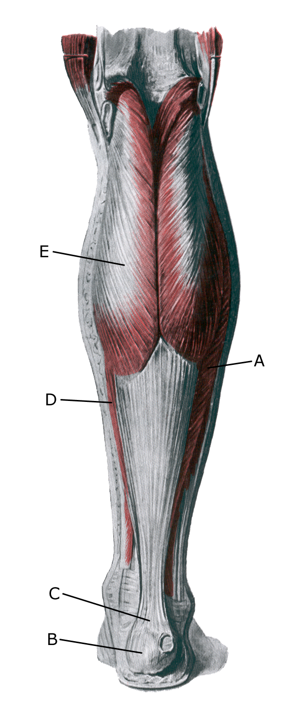
Lower leg from behind:

A. M. soleus
B. Tuber calcanei

C. Tendo calcaneus (Achilles)
D. M. gastrocnemius

Cause of the problem

Partial and total ruptures are a frequent complication of long-term Achilles tendonitis. However, less than half have symptoms before the rupture, but examinations of ruptured Achilles tendon tissue almost always show obvious wear changes (degeneration) in the tendon.

Total ruptures of the Achilles tendon occur most frequently when the athlete lands on the foot (stretching the Achilles tendon) and in the same movement makes a lunge to move forward (contracting the Achilles tendon). This causes a maximal (eccentric) contraction (contraction of the tendon at the same time as it expands), which is common in racket sports (especially badminton).

The changes can be caused by wear and tear changes in the tendon (tendinosis). In recent years, the condition has been called Achilles tendinopathy regardless of whether the cause is inflammation (tendinitis) or wear and tear (tendinosis). Tendinopathy increases the risk of partial and total ruptures.

Symptoms

Sudden onset of pain in the Achilles tendon, often with the sensation and sound of a thud. Many people think they have been kicked from behind. The pain is aggravated by activating the Achilles tendon (walking), pressure on the tendon and stretching the tendon. Often you can feel a defect in the tendon. It is usually impossible to walk on tiptoe.

Examination

In all cases where a pop or sudden shooting pain is felt in the Achilles tendon, you should be examined by an appropriate professional and have a dynamic ultrasound scan performed to diagnose a partial or total rupture of the Achilles tendon.

The vast majority of Achilles tendon ruptures occur 3-6 cm above the heel bone attachment – see ultrasound scan of Achilles tendon with partial rupture, and in a few percent the rupture occurs at the Achilles tendon attachment on the heel bone – see ultrasound scan of inflammation at the Achilles tendon attachment on the heel bone

Treatment

Acute, total ruptures of the Achilles tendon are nowadays primarily treated conservatively (bandaging for 6-8 weeks), with the frequency of surgery decreasing rapidly. However, elite athletes often undergo surgery to get back to elite level faster. Ruptures at the attachment of the Achilles tendon to the heel bone are still recommended for surgery(Cohen D, et al. 2018). Surgery results in fewer re-ruptures, but more side effects. Regardless of the treatment, approximately 9-12 months of injuries must be expected before sports activity can be resumed at the same level.

If surgery has been performed, a rehabilitation guide should be provided by the facility as there may be specific surgical reasons for specific rehabilitation considerations.

Bandage

The foot will usually be bandaged for 6-8 weeks.

Complications

If the progress is not smooth, an ultrasound scan should be performed to rule out re-rupture of the Achilles tendon. Relatively often it will not be possible to return to sporting activity at the same level despite proper treatment and rehabilitation attempts.

Especially

As there is a risk of permanent injury, the injury should be reported to your insurance company

Rehabilitation

Rehabilitation program