Rupture of the muscle on the front of the upper arm

Anatomy

The muscles of the upper arm (brachium) include 3 muscles. Two of the muscles flex the elbow joint (M biceps brachii, M brachialis), while the 3rd muscle (M coracobrachialis) brings the arm towards the body (adduction). The biceps muscle also rotates the hand (suppination).

Shoulder and upper arm muscles from the front:

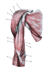
A. M. biceps brachii
B. M. triceps brachii
C. Epicondylus medialis
D. Aponeurosis m. bicipitis brachii
E. M. brachioradialis
F. M. brachialis
G. M. coracobrachialis
H. Tuberculum majus
I. M. deltoideus
J. Acromion
K. Processus coracoideus
L. Clavicula (Collarbone)


Deep muscles on the upper arm

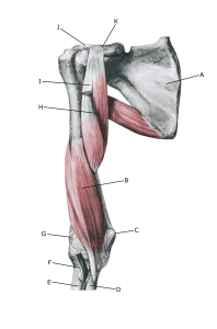
A. Scapula (Shoulderblade)
B. M. brachialis
C. Epicondylus
medialis
D. Ulna
E. Radius
F. Tendo bicipitio brachii (Overcut)
G. Epicondylus lateralis
H. M. coracobrachialis
I. Caput breve (m. bicipitis brachi) (overcut)
J. Caput humeri
K. Processus coracoideus

Cause

When a muscle is suddenly subjected to a load that exceeds the muscle’s strength, a rupture occurs, often accompanied by a haemorrhage. In some cases, the muscle tear (contusion) and haemorrhage occur after a direct blow to the muscle. The vast majority of tears are partial muscle tears. Tears most often occur if the muscle contracts while being stretched (eccentric contraction).

Injuries in muscles can be localised in the muscle tissue itself, in the fascia around/between the muscles or in the tendon part of the muscles, which has a major impact on prognosis (muscles heal much faster than tendons).

Total ruptures are most common in the elderly and are localised to the long head of the biceps tendon at the shoulder joint (caput longum biceps brachii) rupture of the bicepstendor in the shoulder or at the tendon attachment in the elbow rupture of the biceps tendor in the elbow. When the muscle ruptures and especially if it is injured after a direct blow to the muscle, bleeding can occur in the muscle.

In some cases, there may be calcifications corresponding to the bleeding (myositis ossificans) or fluid may be drawn into the bleeding, causing a fluid collection in the muscle (hygroma) complications of muscle haemorrhage.

Symptoms

In mild cases, a localised soreness is felt after the strain (‘muscle strain’, ‘threatening fibre’). In more severe cases, a sudden shooting pain is felt in the muscle (‘partial muscle rupture’, ‘fibre tear’) and in the worst case, a violent pop is felt, after which bending the elbow against resistance is severely impaired (‘total muscle rupture’).

In muscle injuries, the following three symptoms are characteristic: Pain on pressure, stretching (extending the elbow) and activation against resistance (bending the elbow). If there is major bleeding or fluid accumulation in the muscle, you will often feel a firm filling in the muscle.

Examination

The diagnosis is usually made during a clinical examination. Symptoms can be provoked by pressure on the muscle and attempts to activate the muscle (usually bending the elbow or rotating the hand (supination)) against resistance. For more severe pain or difficulty using the arm, an examination by an appropriate professional should be performed to ensure proper diagnosis and treatment.

This can be supplemented with an ultrasound scan, where even minor irregularities in the muscle fibres, bleeding and calcifications can be seen (or an MRI scan).

Treatment

Treatment includes relief from pain-inducing activity, stretching and slowly increasing strength training of the upper arm muscles within the pain threshold.

Localising whether an injury is located in the muscle, in the fascia or in the muscle tendon is of great importance for the speed of rehabilitation. This can often be determined during the clinical examination. If in doubt, ultrasound or MRI scans can pinpoint the localisation of the injury.

Surgery for partial tears in the muscles of the upper arm is rarely indicated. In the case of major haemorrhages or fluid accumulations (hygromas) in the muscle, these can be drained ultrasound-guided, reducing pain and promoting healing.

Complications

If the progress is not smooth, you should be re-examined (again) and consider whether the diagnosis is correct or if there are complications.

In particular, the following should be considered:

Rehabilitation

Rehabilitation program