Anatomy
Behind the medial malleolus, one of the large nerves of the lower leg (tibial nerve) runs down to the foot. The tibial nerve is held in place by a strong tendon (retinaculum musculorum flexorum pedis) in the tarsal tunnel and divides into two nerves: the medial plantar nerve and the lateral plantar nerve. The latter gives off a nerve branch to the heel (Baxter’s nerve). The tibial nerve runs in the tarsal tunnel along with three flexor tendons (tibialis posterior, flexor digitorum longus and flexor hallucis longus) and blood vessels to the foot (arteries) and from the foot (veins).
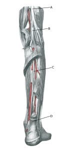
Lower leg from behind:
A. N. ischiadicus
B. N. tibialis
C. N. tibialis
D. Tendo calcaneus (Achillis)
Cause of the problem
The tibial nerve behind and below the medial malleolus can be pinched along with the other nerve branches, causing irritation of the nerve. Numerous causes of nerve irritation have been described, such as shoes that pinch, pinching of the powerful tendon tube in the tarsal tunnel and a fluid collection (ganglion) or tendon and tendon sheath inflammation that presses on the nerve. There is often no explanation for the nerve irritation.
Symptoms
There is a burning, stabbing pain and possibly numbness along the sole of the foot and toes, usually localised to the inner edge of the foot. There may be symptoms from the calf. The pain worsens with walking and running. If the Baxter nerve is affected, there is also pain in the heel, which is often misinterpreted as inflammation of the hollow tendon.
Examination
Usually, general medical examination is sufficient to confirm the diagnosis as the symptoms can be induced by pressing or tapping a finger on the nerve (Tinnel’s sign) behind/under the medial malleolus. The pain subsides (for one hour) when an ultrasound-guided local anaesthetic is applied around the nerve (diagnostic blockade) (Iborra Á, et al. 2023), where even the smaller nerve branches can be affected (Presley JC, et al. 2013).
If in doubt about the diagnosis, ENG (electroneurography) and EMG (electromyography) examination can be performed, which is rarely indicated.
Treatment
Treatment primarily includes relief from pain-inducing activities (running), stretching and slowly increasing rehabilitation within the pain threshold. Wearing high-heeled shoes often relieves the pain.
If there is no steady progress on relief and rehabilitation, treatment can be supplemented with medical treatment in the form of arthritis pills (NSAIDs) or ultrasound-guided injection of adrenal cortical hormone into the tarsal tunnel (Atesok K, et al. 2022). If treatment is ineffective, surgery to expose the nerve can be attempted, but this is rarely necessary (Vij N, et al. 2022).
Complications
If no progress is made, consider whether the diagnosis is correct or if complications have arisen.
In particular, the following should be considered:
- Ligament injury in the ankle joint, inner ligament.
- Concentration of fluid in the joint (traumatic synovitis)
- Slipped disc
- Infections or anything else that can press on the nerve.