Inflammation of the Achilles tendon attachment

Anatomy

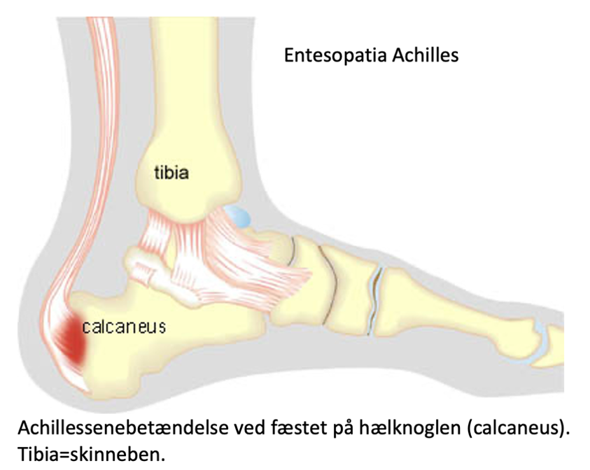
The calf muscle (M Gastrocnemicus) consists of two muscle heads that come together in a broad tendon band that continues into the Achilles tendon. Another large calf muscle (M Soleus) attaches to the front edge of the Achilles tendon and forms part of the Achilles tendon. The Achilles tendon attaches to the heel bone (calcaneus). The Achilles tendon thus consists of the 3 calf muscles (Triceps Surae). At the heel bone, there is a deep mucosal sac at the front (deep bursae tendinis Achilles) and a superficial mucosal sac at the back (superficial bursae subcutanea calcanei) of the Achilles tendon attachment. The bursae reduce the pressure on the Achilles tendon.
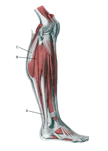
Lower leg:

A. Tendo calcaneus (Achillis)
B. M. soleus
C.M. gastrocnemius

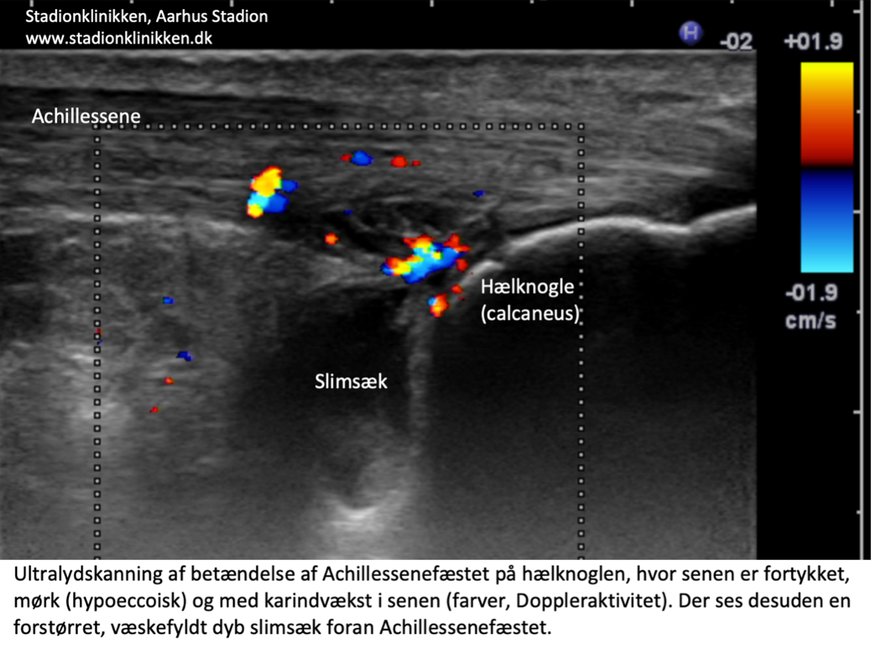
See image of Entesopatia Achilles and Bursitis Achilles

Cause of the problem

“Inflammation” of the Achilles tendon attachment on the heel bone is caused by repeated overloads in the form of running and pushing off. The risk of strain injuries to the Achilles tendon attachment increases with age. Achilles tendonitis at the heel bone attachment increases the risk of (partial) rupture of the Achilles tendon.

Inflammation of the deep bursa in front of the Achilles tendon attachment often occurs simultaneously with strain-related Achilles tendonitis at the heel bone attachment.

In some cases, Achilles tendonitis at the heel bone attachment is part of a rheumatic disease (axial spondyloarthritis and psoriatic arthritis)

The changes can be caused by wear and tear changes in the tendon (tendinosis). In recent years, the condition has been called Achilles tendinopathy regardless of whether the cause is inflammation (tendinitis) or wear and tear (tendinosis). Tendinopathy increases the risk of partial and total ruptures.

Symptoms

Pain when activating the Achilles tendon (running and pushing off) and pressing on the tendon attachment on the heel bone. In contrast to the soreness of Achilles tendonitis, the soreness is localized all the way down to the attachment on the heel bone (enthesis). With Achilles tendonitis in front of the Achilles tendon attachment, there is also tenderness on pressure just in front of the heel bone attachment. Symptoms can last for several years in some cases.

Examination

Diagnosis in mild cases, where the pain is slow onset without sudden worsening, is made during a clinical examination. In cases where a pop or sudden shooting pain is felt at the tendon attachment, you should be examined by an appropriate professional and have a dynamic ultrasound scan performed to diagnose a (partial) rupture of the Achilles tendon attachment.

‘Inflammation’ at the Achilles tendon attachment on the heel bone is caused by repeated overloads in the form of running and pushing off. The risk of strain injuries to the Achilles tendon increases with age. Achilles tendonitis at the heel bone attachment increases the risk of (partial) rupture of the Achilles tendon (=FOD-16).
Inflammation of the deep bursa in front of the Achilles tendon attachment often occurs simultaneously with strain-induced Achilles tendonitis at the heel bone attachment.
In some cases, Achilles tendonitis at the heel bone attachment is part of a rheumatic disease (axial spondyloarthritis and psoriatic arthritis)
See ultrasound scan of the Achilles tendon attachment on the heel bone.

Treatment

Treatment for overuse Achilles tendonitis at the heel bone attachment primarily consists of relief from the pain-inducing activity (running), stretching and slowly increasing strength training within the pain threshold. If treatment is started quickly, the Injury can sometimes heal in a matter of weeks. If the pain has been present for several months and ultrasound scans show thickening and changes in the Achilles tendon attachment, a rehabilitation period of several months/half years is often required.

Special emphasis is placed on strength training by activating the tendon while stretching it (eccentric training) or slow heavy strength training(Johannsen F, et al. 2018). If relief and rehabilitation are not successful, rehabilitation can be considered supplemented with medical treatment in the form of arthritis pills, NSAIDs or injection of adrenal cortex hormone(Johannsen F, et al. 2018) into the deep bursa in front of the Achilles tendon attachment on the heel bone. Injecting into the deep mucosal sac avoids troublesome contraction of the fatty tissue (fat atrophy), which is a possible complication of the injection and can cause discomfort from the heel cup of the shoe.

As adrenal cortex hormone injection is always part of a long-term rehabilitation of a very serious, chronic Injury, it is crucial that the rehabilitation process lasts (at least) six months to reduce the risk of relapse and rupture. Of course, after a long-term injury, the tendon cannot withstand maximum strain after only a short-term rehabilitation period.

Six months of rehabilitation before maximum load (jumping, sprinting) is allowed is not unusual. See General, Chronic tendonitis.

It is important that the heel cup of the shoe does not press on the Achilles tendon/sole and aggravate the discomfort. A pressure-relieving insert in the heel cup can reduce the pressure on the tendon and bursa.

Acute total ruptures of the Achilles tendon attachment are usually treated surgically with subsequent bandaging for 6-8 weeks(Cohen D, et al. 2023).

Complications

If the progress is not smooth, a (repeat) ultrasound scan should be performed to rule out (partial, progressive or total) rupture of the Achilles tendon attachment.

Rehabilitation

Rehabilitation program