Anatomy
Ledfladerne er beklædt med en få mm tyk bruskbelægning, der tjener til at nedsætte belastningen på ledfladerne.
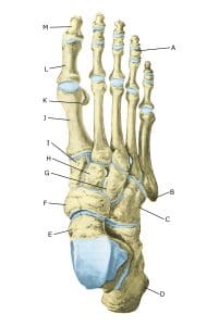 Foot from above:
A. Phalanx media
B. Tuberositas ossis metatarsalis V
C. Os cuboideum
D. Calcaneus
E. Talus
F. Os naviculare
G. Os cuneiforme laterale
H. Os cuneiforme intermedium
I. Os cuneiforme mediale
J. Os metatarsalei
K. Os sesamoideum
L. Phalanx proximalis
M. Phalanx distalis
Cause
Cartilage damage in the articular surfaces can occur in several ways. Severe twisting of the joint, where the articular surfaces collide, can cause localised cartilage destruction. In other cases, a piece of bone can be rejected from a joint surface for no known reason (osteochondritis dissecans). Bone fragments can be moved around the joint (joint mouse) and become trapped in the joint, causing locking so that the joint cannot move.
Repeated (over)stress can also damage cartilage and result in osteoarthritis. Cartilage changes in joints can sometimes cause irritation of the synovium (synovitis), resulting in increased fluid accumulation in the joints of the foot with swelling, restriction of movement and pain in the joint.
Osteoarthritic changes in the joints are most common after fractures, repetitive ligament injuries to the external ligaments of the ankle joint, internal ligament injuries to the ankle joint and especially common after fractures in the articular surface.
Most commonly affected are the ankle joint, the joints between the metatarsal bones of the metatarsus and the metatarsophalangeal joint of the big toe stiffness of the big toe (hallux rigidus)
Symptoms
There is pain in the joint when moving with strain. In addition, there can sometimes be ‘inflammation’ of the synovial membrane (synovitis), which causes fluid accumulation and increasing pain in the joint.
Examination
The diagnosis is made through a clinical examination, but it is often necessary to supplement this with imaging in the form of X-rays, ultrasound or MRI scans. In some cases, a binocular examination of the joint (arthroscopy) is necessary.
Treatment
Treatment includes relief from pain-inducing activities. Gentle rehabilitation within the pain threshold can begin immediately. If the discomfort is triggered from the toes or metatarsal, a stiff sole will reduce stress on the joint. Joint mobility and muscle strength should be maintained without provoking the joint to cause further pain and swelling.
Conditioning can be done by cycling or swimming. If the swelling in the joint does not subside despite relief, it can be supplemented with medical treatment in the form of arthritis pills (NSAIDs) or possibly fluid drainage and ultrasound-guided injection of adrenal cortex hormone into the joint.
There is no treatment that can restore the damaged cartilage, which has little ability to heal. In some cases, surgery is indicated (Walther M, et al. 2023), especially if there is a possibility to fix a rejected bone/cartilage piece (osteochondritis dissecans) (Bruns J, Habermann C, Werner M. 2021).
Complications
Large cartilage injuries located on the weight-bearing parts of the (ankle) joint are a serious sports injury and often lead to the cessation of sport.
In particular
As there is a risk that the injury may cause permanent damage, you should report the injury to your insurance company.