Anatomy
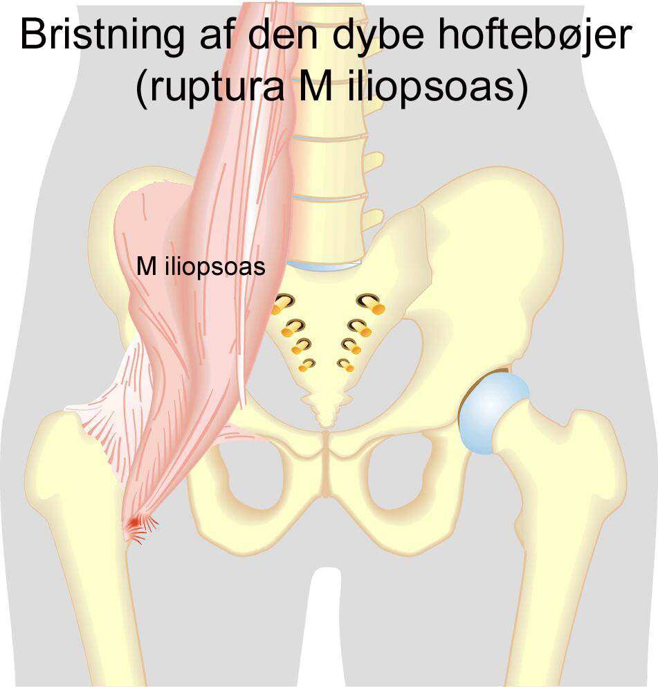
The deep hip flexor (M iliopsoas) consists of two muscles. The psoas muscle originates from the lumbar vertebrae and the iliacus muscle originates from the inside of the hip bone. The two muscles fuse together and both attach to the inside of the femur (trochanter minor). The iliopsoas is the strongest flexor muscle of the hip joint.
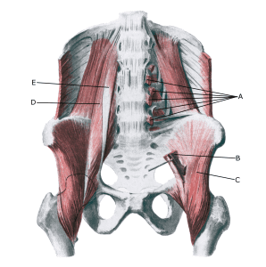
Pelvis from the front:
A. Origines m. psoatis
B. M. psoas major
C. M. iliacus
D. M. psoas major
E. M. psoas minor
Cause
When a muscle is subjected to repeated strain that exceeds muscle strength (jumping, kicking), damage occurs to the muscle and muscle tendon. The repeated overloads can result in chronic “inflammation” and small tears that weaken the tissue and in the worst case result in a total rupture.
Symptoms
The pain is localised in the middle of the groin and above the hip joint and may radiate slightly up the abdomen, down the inside of the thigh and into the lower back. In mild cases, a localized soreness is felt at the start of exercise, but the symptoms can often fade after a thorough warm-up and return after the sporting activity has stopped (“muscle strain”, “threatening fiber”, “tendonitis”).
In more severe cases, a sudden shooting pain is felt in the muscle (“partial muscle rupture”, “fibre rupture”) and in the worst case, a violent snap is felt, making it impossible to use the muscle such as climbing stairs (“total muscle rupture”).
In muscle injuries, the following three symptoms are characteristic: Pain when pressing, stretching and activating against resistance. In rare cases, the hemorrhage can become so severe that it pinches the nerve to the leg (femoral nerve) with increasing pain, loss of strength and symptoms down the leg.
Examination
The diagnosis is usually made on clinical examination alone, with localised pressure soreness on the muscle that worsens with stretching and muscle contraction. If the injury occurs suddenly with the sensation of a pop and decreased function of the muscle (flexion of the hip), a medical examination is advisable. If there is any doubt about the diagnosis, an ultrasound or MRI scan.
Treatment
Treatment primarily includes relief from pain-inducing activity, stretching and graduated rehabilitation within the pain thresholdAnderson CN. 2016). If no progress is made despite regular rehabilitation, rehabilitation may be supplemented with injection of adrenal cortical hormone around the inflamed (inflamed) part of the iliopsoas, accompanied by a rehabilitation period of several months to reduce the risk of recurrence and rupture.
Surgery is usually not indicated, but may be attempted in special cases. Surgery for total ruptures is not indicated (Anders A, Vitale K. 2022). Very large haemorrhages can be drained ultrasound-guided.
Complications
If no progress is made, you need to consider whether the diagnosis is correct or if there are complications:
In particular, the following should be considered: