Anatomy
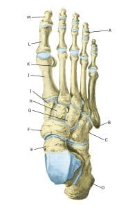
The foot below
A. Ossa sesamoidea
B. Talus
C. Calcaneus
Cause
Repeated overextension of the ankle joint (plantar flexion) or maximum flexion of the ankle joint (dorsiflexion) can cause damage to the bones due to impact or excessive strain on the ligament attachments at the front and back of the ankle joint.
This leads to the formation of bony spurs (exostoses), which can cause soft tissues from the ligaments and joint capsule to become pinched (impingement) between the bony spurs when the ankle joint is moved. If the impingement occurs at the front of the ankle joint, it is called anterior footballer’s ankle, and if the impingement occurs at the back of the ankle joint or subtalar joint, it is called posterior footballer’s ankle (K. Lavery, et al. 2019). Approximately 5% of people have an extra (accessory) bone at the back of the ankle (os trigonum), which can contribute to the impingement; this is known as os trigonum syndrome. 2% have an os trigonum at the back of both ankles. In some cases, an enlargement of a bony prominence on the talus is observed, known as the Stieda process.
Footballers, volleyball players, rugby players, javelin throwers, gymnasts, dancers and athletes who perform a lot of jumping movements are at greater risk of spraining their ankles. Previous ligament injuries in the ankle joint also increase the risk due to scar tissue formation, which is more easily pinched between the bony prominences.
Symptoms
Pain at the front or back of the ankle joint, particularly when bending and stretching to the extreme positions (plantar and dorsal flexion), such as when kicking a ball or pushing off (at the back). Even everyday movements (climbing stairs) can trigger the pain. There may be swelling following physical activity. Some people experience a ‘clicking’ sensation during plantar and dorsal flexion of the ankle joint.
Examination
Diagnosen kan sædvanligvis stilles alene ved klinisk undersøgelse, hvor der er karakteristisk ømhed ved bevægelse og tryk forpå eller bagpå ankelleddet. Det kan være nødvendigt at supplere med røntgen og ultralydskanning, mens MR- og CT-skanning sjældent er nødvendigt. Ved ultralydskanning er det muligt at se mindre knogleforandringer, hævelse og inflammation af de afklemte bløddele.
Treatment
Treatment primarily involves avoiding activities that trigger pain, as well as exercises aimed at improving mobility in the ankle joint. In cases of anterior ankle pain, heel inserts or heel lifts may provide relief. If the pain persists despite rest and rehabilitation, medical treatment may be added in the form of anti-inflammatory tablets (NSAID) or ultrasound-guided injections of corticosteroids around the tender soft tissues. Around 75% become symptom-free within 3–6 months.
If pain persists and does not subside despite rest and medical treatment, further investigations, including arthroscopy, and surgery may be indicated, during which bone spurs (and possibly the os trigonum or Stieda) and trapped tissue are removed (NSB Mansur. Et al. 2024 , X Chin, et al. 2023).
Complications
If the condition does not improve steadily, one should consider whether the diagnosis is correct or whether complications have arisen:
Bone fracture in the forefoot
Tendon synovitis
Joint cartilage damage, osteoarthritis
Concentration of fluid in the joint
Ligament injury in the ankle joint, outer ligament
Ligament injury in the ankle joint, inner ligament