Anatomy
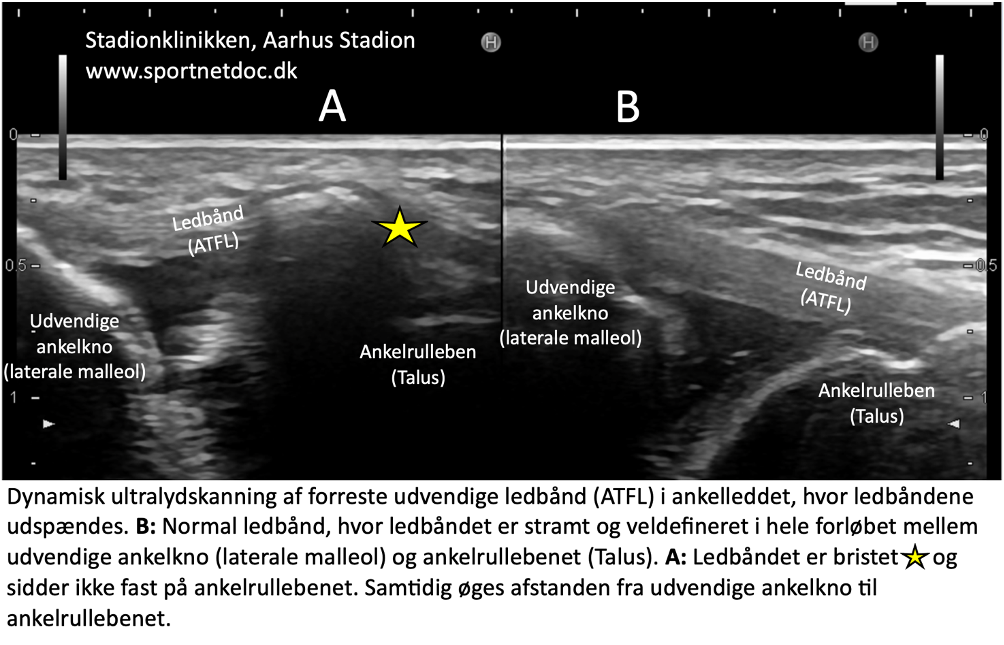
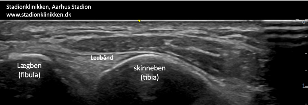
The ankle joint is stabilised by a joint capsule that is reinforced internally (medially) by a wide, fan-shaped ligament (ligamentum deltoideum) and externally (laterally) by 3 ligaments: an anterior (ligamentum talofibulare anterius), a middle (ligamentum calcaneofibulare) and a posterior (ligamentum talofibulare posterius). The ligaments stabilise the ankle joint, especially when twisting, running with changes in direction, etc. The tibia and fibula are held together in the ankle joint by 2 ligaments (ligamentum tibiofibulare anterius and posterius).
Ankle joint, External
A. Ligamentum mediale/deltoideum
B. Calcaneus
C. Talus
D. Tibia (Shin bone)
Cause of the problem
Rupture of the lateral collateral ligaments of the ankle joint is one of the most common sports injuries and occurs when the foot wiggles (supination), overstretching and rupturing the outer ligaments. In mild cases it is called a sprain/strain and in severe cases a complete or partial rupture/tear.
The anterior talofibular ligament (ATFL) ruptures most frequently. The middle ligament (calcaneofibular ligament) ruptures in 20% of cases where the anterior ligament ruptures.
The posterior ligament usually only ruptures in severe ankle injuries with fractures or dislocations. There are often secondary injuries associated with ligament ruptures, including rupture of the ligaments that hold the tibia and fibula together (syndesmosis rupture), fluid accumulation in the joint (traumatic synovitis), tendonitis, rupture of ligaments in the metatarsal, ankle fractures, cartilage damage inside the ankle joint (major cartilage lesions are seen in 7% of sprains) and damage to the subtalar joint (the joint between the heel bone (calcaneus) and ankle roll bone (talus)).
In some cases, ankle ligament ruptures are complicated by inflammation of the joint capsule around the ankle joint (capsulitis).
Symptoms
Pain on and below the outer ankle bone (malleolus lateralis), swelling and discolouration due to bleeding and pain when walking.
Examination
Diagnosis is made by clinical examination, where in mild cases (sprains) there is minimal swelling and no discomfort with normal walking, but always pressure tenderness at the anterior cruciate ligament attachment (ATFL). The amount of swelling is not a measure of the extent of the injury. Pronounced swelling or pain should be examined by an appropriate professional to rule out ankle fracture and rupture of the ligaments between the tibia and fibula (syndesmosis rupture).
If a fracture is suspected, an X-ray of the ankle joint should be taken. The Ottawa ankle rule provides guidance on when an X-ray should be taken (Yolanda E Gomes et al 2022).
Previous vigorous twisting of the ankle joint in the acute stage to assess the degree of looseness is no longer indicated as this does not affect the choice of treatment.
Ultrasound scanning can produce external ligaments – see ultrasound scan here and the ligament (lig. tibiofibulare ant. inf.) between the tibia and fibula (syndesmosis) – see ultrasound scan here and inflammation in capsulitis (Colò G, et al. 2023). Ultrasound scanning is well suited to see small bone tears at the ligament attachments.
Dynamic ultrasound scanning can assess the looseness of the ligaments (the ligament is stressed during simultaneous ultrasound scanning to see if the joint gap increases as a sign of ligament rupture – Heitz PH, et al. 2023).
Watch the video here: Ultrasound scan of the lateral collateral ligaments of the ankle while moving (supinating) the ankle joint. This increases the distance between the lateral malleolus (left) and the calcaneus (right), indicating that the lateral collateral ligament is not functioning. This proves that the ligament has ruptured
Treatment
The treatment of ligament injuries today is conservative (rehabilitation). In the past, many people underwent surgery and bandaging, which has largely been abandoned for uncomplicated ligament ruptures. In the case of repeated ankle sprains and syndesmosis ruptures with significant looseness, surgery is often indicated (Colò G, et al. 2023).
Bandage
During rehabilitation, tape (or different types of ankle braces) is often used when running on uneven surfaces or running with rapid changes of direction. The ligaments contain nerve cells (proprioreceptors) that send information to the brain about the position of the ankle joint.
Information is sent from the brain to the muscles, which are activated to keep the ankle joint in the correct position. When the ligaments are damaged, these nerve pathways don’t function optimally, increasing the risk of twisting the foot again.
The primary function of the tape is to stimulate the small nerve cells (proprioreceptors) in the skin so that they can ‘substitute’ for the temporarily damaged nerve cells in the ligaments. The function of the tape is thus not just a purely mechanical ‘stabilisation’ of the ankle joint – See tape
In some cases, certain types of bandages around the ankle joint can be beneficial for 5-6 weeks. Specialised bandages have been shown in some studies to reduce the risk of ligament re-injury (Zwipp H. 2023).
Complications
If the progress is not smooth, you should consider whether the diagnosis is correct or if there are complications:
In particular, the following should be considered:
- Rupture of ligaments between the tibia and fibula (syndesmosis rupture),
- Bone fracture
- Periosteal tears (avulsio),
- Tendon luxation, outer ankle knuckle (peroneus luxation)
- Tendon synovitis
- Concentration of fluid in the joint (traumatic synovitis)
- oint cartilage damage, osteoarthritis (osteochondral lesion)
- Rupture of the Achilles tendon
10-15% of athletes with injuries to the external ligaments of the ankle continue to have discomfort 1 year later, probably due to insufficient rehabilitation before resuming full sports activity.
Especially
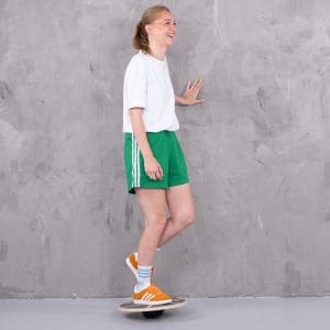
Rocking board exercises are important in both rehabilitation and prevention (Al Attar WSA, et al. 2023). Rocking board exercises should be performed regularly for the rest of the active sports career as a preventive measure if there is a history of ankle ligament injuries.
Start by standing on both legs on the seesaw and support with your hands on the wall. Gradually release the support with your hands and finally practice with support on one leg only.
You can brush your teeth morning and evening on the rocker board (Wang F, et al. 2023).